Next tests schedules


Oocytes

The morphology of the oocyte can be described by its subcellular phenotypic constituents: the zona pellucida, the perivitelline space, the polar bodies and the cytoplasm. There are infinite variations of these structures and the best way to describe them is through micrographic pictures.

Zona pellucida (ZP)

The thickness of the ZP may vary from <15 µm to > 20 µm. It may also present a septum or a dark color.

EX_ZP_001.jpg

ZP_thickness_age.jpg

Perivitelline space (PVS)

The perivitelline space (PVS) may be large or nearly absent. It may contain fragments.

EX_PVS_001.jpg

First polar body (PB1)

PB1 is considered as normal when it is oval and intact, with a smooth or rough surface. Fragmented or giant PB is considered as abnormal.

EX_PB_001.jpg

Cytoplasmic inclusions and clusters

  • vacuoles: fluid-filled refringent round cavities. They may be isolated or numerous.
  • refractile body: inclusions of different sizes containing lipofucsin
  • endoplasmic reticulum (SER) cluster: appears as a flat elliptical disk in the centre of the ooplasm
  • dark cluster: a cluster of organelles that appears as a dark area in the ooplasm

EX_INC_001.jpg

Cytoplasmic granularity gradients

A granulated area is frequently observed in the animal (PB1 region) or vegetal poles (opposite to the animal pole) but also in a meridional region extending from the vegetal to the animal pole.

EX_GRAN.jpg

Parameters of the EQC test

The EQC test will present you several possible answers for the various characteristics of an oocyte. You can make only one choice. Although you may not be used to grade the oocytes in your day-to-day activity, grading is a convenient way of communicating oocyte characteristics both to colleagues and medical staff. In our view, this is an important parameter to monitor in an External Quality Control scheme.


Zona pellucida

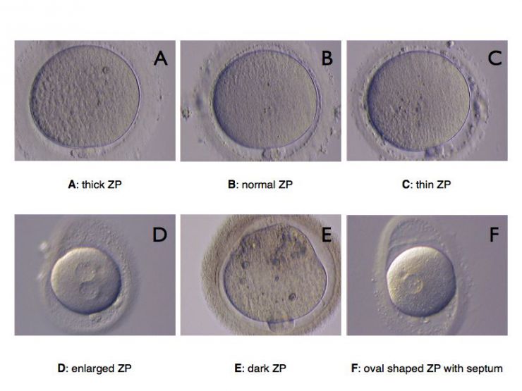
The zona pellucida (ZP) may vary in thickness and rigidity. Some patients have clearly thinner ZP than others. It is usually a characteristic associated with the patient more than with the oocyte. Although thickness may vary slightly around the oocyte, the following categories can be defined.


Parameters Morphological aspects
Thin the ZP is less than 15 µm
Thick the ZP is greater or equal to 25 µm
Normal the ZP is around 20 µm (15-25)
Abnormal the ZP is quite irregular both in thickness and in shape

Perivitelline space

The perivitelline space surrounds the ooplasm. It is generally clearly visible, at least in the vicinity of the polar bodies. It may contain fragments.


Parameters Morphological aspects
Small the ooplasma is adherent to the ZP and the perivitelline space is almost inexistent
Large the ooplasma is clearly retraced away from the ZP leaving a clearly identifiable space all around the cytoplasm
With fragments presence of clearly identifiable fragments
Normal the ooplasm is slightly retracted away from the ZP at least in the vicinity of the polar body


Polar body

The polar body appears when the first meiotic division has been completed. It consists of a small cell within the PVS, close to the oocyte nucleus, and may undergo a second division. It usually rapidly degenerates and appears as a fragmented structure.


Parameters Morphological aspects
Unclear or absent the polar body may not always be clearly identifiable, depending on how the oocyte is oriented. It is absent in a metaphase I oocyte (before the first meiotic division)
Intact clearly identifiable smooth round/oval cell lying close to the oocytes
Fragmented the polar body material is irregular in shape and show clear signs of disorganization
Giant usually intact, giant polar bodies a clearly recognizable, they may achieve size of about half of that of an oocyte


Cytoplasm

Texture

The cytoplasm of the oocyte exhibits usually a granular texture. Depending on the oocyte, this texture may appear homogeneous or exhibit a gradient in the granulated texture. In some cases, it may contain a dark cluster and exhibit sign of fragmentation.


Parameters Morphological aspects
Fragmented the cytoplasm shows signs of fragmentation or appear as an undefined mass with absent or degraded plasma membrane
Homogeneous the cytoplasmic material appears homogeneously distributed with little signs of granulation
Polarised zones more or less granulated are visible as two opposing hemispheres
Dark cluster a dark zone is clearly identifiable inside the cytoplasm


Inclusions

The cytoplasm may contain several remarkable structures.

Parameters Morphological aspects
Vacuole one large or several small vacuoles bounded with a clearly identifiable membrane
SER cluster smooth endoplasmic reticulum appearing as clear spherical zones inside the cytoplasm. The boundaries of these structures may not be easily identified as in the case of vacuoles
Refractile body Refractile bodies constitute one of the main morphological abnormalities in human oocytes and are made of autofluorescing lipofucsin (appear yellowish)
None absence of the above-mentioned structures

Effects of various phenotypes on embryo development and implantation

The influence of the different oocyte phenotypes on embryo development and implantation is still controversial. Some dysmorphic morphological features may represent phenotypic variability without any consequences on development.


Parameters Effect on implantation
ZP abnormality Controversial
PVS size Controversial
PVS fragments Controversial
PB morphology Controversial
SER Detrimental if severe
Vacuoles Detrimental if severe
Refractile bodies Detrimental if severe
Dark clusters Detrimental if severe


Reference

Balaban B and Urman B. Effect of oocyte morphology on embryo development and implantation. RBMonline 12: 608-615, 2006